Visual Differences: How to Instantly Tell Decorticate from Decerebrate Posturing
 By Sarah Jenkins
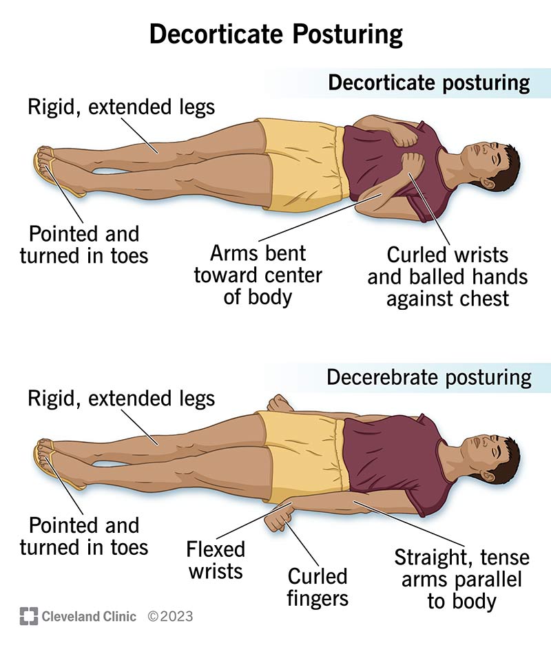
Emergency medical services, trauma surgeons, and neurointensivists use specific physical and clinical metrics to differentiate abnormal motor activity. The clinical picture shifts considerably across physical presentations, underlying structural drivers, and prognostic outcomes.
| Clinical Feature | Decorticate Posturing | Decerebrate Posturing |
|---|---|---|
| Upper Limb Position | Flexion at elbows, wrists, and fingers; held tight to chest | Rigid extension, internal adduction, forearms fully pronated |
| Lower Limb Position | Rigid extension with internal rotation and plantar flexion | Rigid extension with severe plantar flexion and inward turning |
| Anatomical Level | Above the midbrain / Rostral to red nucleus (cortex, thalamus) | Midbrain to upper pons / Caudal to red nucleus |
| Active Motor Pathway | Rubrospinal (arms flex) + Vestibulospinal (legs extend) | Vestibulospinal and reticulospinal tracts unopposed |
| Glasgow Coma Motor Score | Score of 3 (Abnormal flexion) | Score of 2 (Abnormal extension) |
| Primary Etiologies | Diffuse axonal injury, cortical stroke, early uncal herniation | Central/tonsillar herniation, pontine infarct, brainstem hemorrhage |
| Immediate Reversibility | Moderate; high urgency to avoid downward herniation | Low to critical; demands emergent surgical or osmotic decompression |
Sarah Jenkins is a veteran tech journalist with over 12 years of experience covering artificial intelligence, mobile innovations, and digital ethics. Her insights have appeared in leading technology publications worldwide.
Tags:
posture decorticate decerebrate
