Mapping the Gut Immune System: Where Peyer’s Patches Actually Reside
 By David Miller
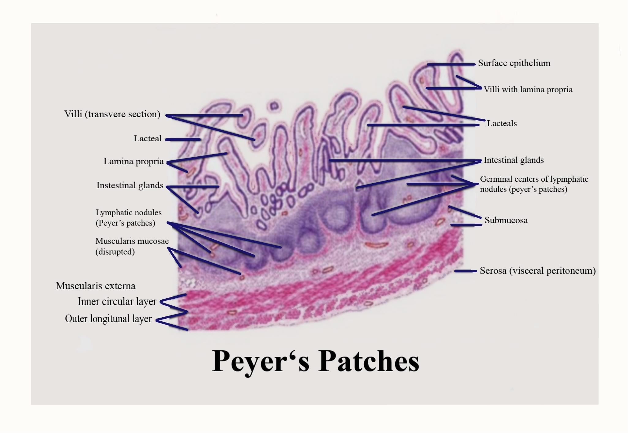
To understand where these lymphoid organs sit, anatomists look at how gut-associated lymphoid tissue is organized across the three segments of the small intestine.
The duodenum harbors the lowest bacterial load because gastric acid and bile wash through regularly. True macroscopic Peyer's patches rarely develop here. Instead, the duodenum relies on isolated lymphoid follicles (ILFs), tiny microscopic solitary nodules scattered across the mucosa. Very rarely, deep biopsies capture solitary follicles that cluster into an immature aggregate, but classic elongated patches do not form in the proximal duodenum.
Moving into the jejunum, the landscape changes. The proximal jejunum features high circular folds designed for nutrient absorption. As one travels distally toward the ileum, the plicae circulares begin to shrink, while aggregated lymphoid follicles start to emerge. In healthy young adults, roughly 15% to 20% of the body's total Peyer's patch inventory sits within the distal jejunum. These jejunal patches tend to be rounder, shorter, and separated by wide stretches of mucosa.
The ileum represents the primary operational base for aggregated follicles. Here, especially in the distal third and terminal ileum, patches reach lengths of several centimeters, visible to the naked eye on the antimesenteric border (opposite the mesenteric attachment). Histological surveys show that an adolescent possesses between 150 and 250 distinct Peyer’s patches across the small intestine, with roughly 80% to 85% packed into the ileum.
| Small Intestine Segment | Peyer’s Patch Distribution | Follicle Architecture | Primary Immunological Task |
|---|---|---|---|
| Duodenum | Extremely rare to absent (<1%) | Isolated lymphoid follicles (ILFs); tiny micro-aggregates | Baseline baseline chemical defense; local secretory IgA |
| Jejunum | Moderate (15%, 20%, mostly distal) | Small, discrete circular patches; fewer germinal centers | Surveillance of partially digested food antigens and commensals |
| Ileum | Dense (80%, 85%, peaks at terminal ileum) | Large, confluent oval plaques (up to 10 cm); dense germinal zones | Massive antigen sampling; high-volume IgA class switching |
David Miller brings 15 years of experience in global economics, personal finance strategy, and market dynamics. He specializes in turning complex economic trends into actionable insights for everyday readers.
